How Sialendoscopy is done?
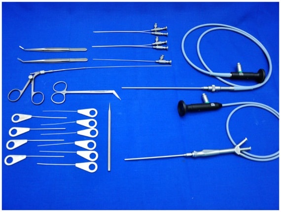
Local anesthesia is infiltrated around the papilla of the salivary duct in order to make the papilla taut and also to reduce the bleeding around the papilla. Smallest size probe (00 size) is introduced into the papilla. This papilla is then dilated with serial dilators of increasing size. If one find difficulty in identifying the papilla one may use microscope for this purpose or one may massage the gland to identify the saliva coming out of papilla. Then the diagnostic sialendoscope is introduced along with the sheath with continues irrigation so that while introducing the sialendocope the duct remains open. If there are any mucous plugs in the duct, they also get washed away with this irrigation. Once the inspections of all the branches of the duct is done, and if any pathology is seen then sialendoscope with the therapeutic sheath is introduced and treatment is done accordingly.
Opening Hours - Sir Ganga Ram Hospital
- Monday - Friday
6:00 PM - 8.00 PM
Opening Hours - Clinic
- Monday
10:30 AM - 12:30 PM
- Wednesday
10:30 AM - 12:30 PM
- Saturday
10:30 AM - 12:30 PM
Patient Testimonials

Piyush Goel
IT Professional
I was suffering from severe cough, cold and headache. I consulted with so many doctors, but did not get relief. My friend told me about Dr. Satinder and I consulted with him. He gave five days medicine for these symptoms. Surprisingly, I got relief in two days only. I am thankful for his great treatment that he had given to me. Thanks a lot.

Ravinder Pal Singh Sethi
IT Professional
I can't explain how much pain I was going through due to the mump at right side of my face. I got really scared as ear & throat both were paining. My boss suggested Dr. Satinder Singh, ENT specialist. Really Thanks to him for such a kind support and treatment. Within a day I was relived from the pain.

Few days back, got ulcers on the throat due to which unable to even inhale or eat or drink anything. Come to Dr. Satinder and really it was really a safest and quickest treatment i got and started normal diet within 3 days.